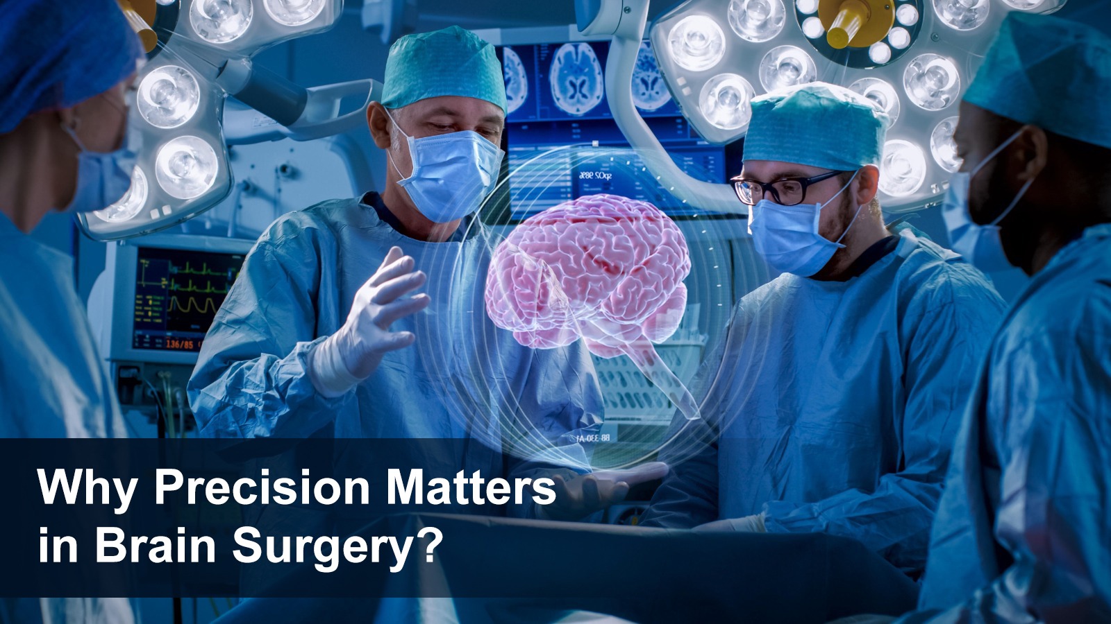
Why Precision Matters in Brain Surgery: The Role of the Neuromicroscope
In the field of brain surgery, efficiency is essential. A fraction of a millimeter can be the difference between restoring health and risking vital function. That's why modern neurosurgeons rely on a game-changing tool: the neuro microscope.
These advanced systems, like the ones from ZEISS TIVATO 700 neuromicroscope, are transforming patient outcomes by offering unmatched clarity, depth, and control. Let’s explore why neuromicroscopes are essential in today’s neurosurgical procedures—and how they’re helping patients get safer, better results.
What is a Neuromicroscope?
A neuro microscope (also known as a neurosurgical microscope) is a high-powered, high-resolution optical system used in delicate brain and spinal surgeries. It not only magnifies tiny structures inside the brain and spine, but helps to differentiate between normal and abnormal tissues thus allowing surgeons to operate with extreme accuracy.
These aren't ordinary microscopes. A high-resolution neurosurgical microscope offers:
• 3D depth perception
• LED or xenon illumination
• Autofocus and motorized zoom
• Ergonomic, hands-free control
• Integrated imaging and recording
Modern neurosurgery microscope systems give surgeons a clear, magnified view of the operating field, helping avoid damage to critical nerves and tissues.
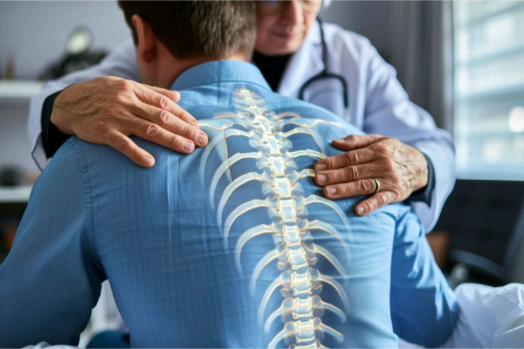
Why Precision Matters in Brain and Spine Surgery
The brain is a delicate organ, home to billions of neurons that control everything from speech and memory to movement and breathing. Even a tiny error can result in serious complications. Here’s why precision is non-negotiable:
• Microscopic surgical areas: Tumors, aneurysms, and malformations are often millimeters in size.
• High-risk zones: Many surgeries involve areas near critical structures like the brainstem or spinal cord.
• Irreversible damage: Unlike other tissues, damaged brain cells don’t regenerate easily.
Differentiation between normal and abnormal tissues
This is where microscope-assisted neurosurgery becomes life-saving.
Benefits of Using a Neurosurgical Microscope System
Using a neurosurgery microscope offers multiple benefits to both the surgical team and the patient:
1. Greater Surgical Accuracy
• Enhanced visualization of blood vessels and nerves
• Clear distinction between healthy and abnormal tissue
2. Minimally Invasive Surgery
• Smaller incisions
• Reduced trauma to surrounding tissue
• Faster healing
3. Reduced Risk of Complications
• Avoids unintentional nerve damage
• Precise control minimizes bleeding
4. Better Long-Term Outcomes
• Higher success rates
• Improved recovery
• Lower risk of recurrence
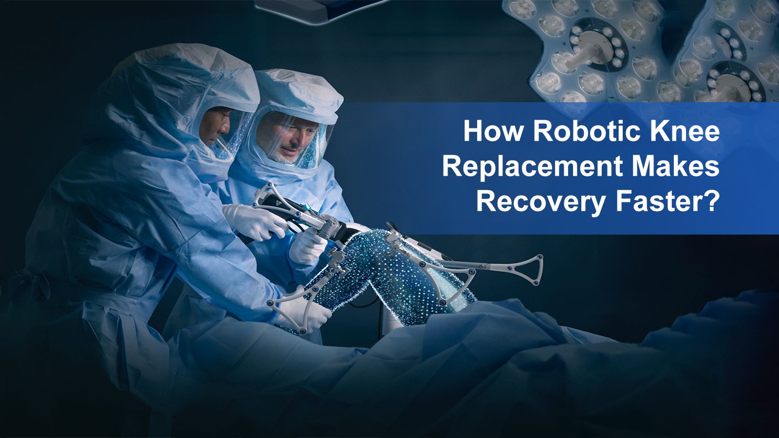
Microscope-Assisted Spine Surgery
Neuromicroscopes are not just for the brain. Operating microscopes for spine surgery are crucial for treating conditions like herniated discs, spinal tumors, and nerve compression. In these procedures:
• Surgeons use a surgical operating microscope to navigate narrow spinal spaces
• Enhanced visualization reduces damage to nerve roots
• Precision helps preserve spinal stability
Trusted Technology: ZEISS TIVATO 700 Neuromicroscope
At Techno India DAMA Hospital, our neurosurgical team uses the advanced ZEISS TIVATO 700 neuromicroscope, a state-of-the-art system designed to redefine surgical precision and performance.
This cutting-edge neurosurgical microscope offers:
• Ultra-clear, high-definition optics for unmatched visualization
• Integrated 4K video and digital connectivity for enhanced surgical planning and documentation
• Advanced ergonomics and intuitive control to support long, complex procedures with surgeon comfort
• Real-time visualization and teaching capabilities for intraoperative collaboration
The ZEISS TIVATO 700 sets a new benchmark in neurosurgical microscope systems, helping our surgeons achieve superior outcomes with enhanced precision, safety, and confidence.
Real-Life Applications of Neurosurgery Microscopes
Common procedures assisted by an operating microscope include:
• Brain tumor removal
• Aneurysm clipping
• Arteriovenous malformation (AVM) surgery
• Trigeminal neuralgia decompression
• Cervical or lumbar spine decompression
• Microvascular decompression
Each of these procedures involves intricate anatomy that’s only visible under a neuro microscope.
Choose the Best in Neurosurgical Care
If you or a loved one requires brain or spinal surgery, choosing a hospital that uses the latest neurosurgical microscope system can make all the difference.
At Techno India DAMA Hospital, our expert neurosurgeons are equipped with advanced microscope systems from ZEISS TIVATO 700 neuromicroscope, delivering world-class care with precision and compassion.
Learn more or book a consultation today
Final Thoughts
The evolution of microscope-assisted neurosurgery has redefined what’s possible in brain and spine surgery. With the help of a neuro microscope, surgeons can work smarter, safer, and more effectively—giving patients better chances at full recovery.
In an area where every millimeter counts, tools like the neurosurgery microscope, surgical operating microscope, and high-resolution neurosurgical microscope aren’t just optional—they’re essential.
Choose precision. Choose expertise. Choose Techno India DAMA Hospital.
For more information about our neurosurgery services and the advanced tools we use, visit:
https://technoindiahealth.com


.webp)
.webp)